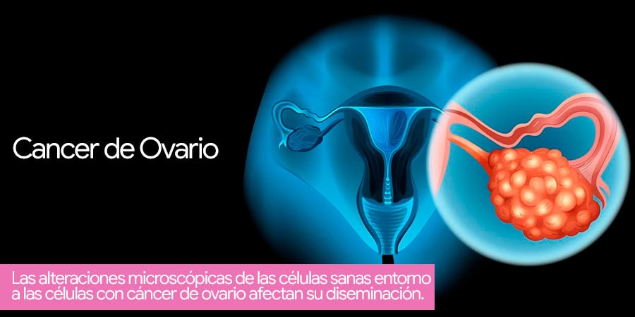
Microscopic changes in healthy cells around cells with ovarian cancer affect their spread
Biophysics researchers at the University of Wisconsin have applied the concept of topological defects to investigate the spread of ovarian cancer cells. Using an experimental model, the researchers found that these defects, or disruptions in normal cell design, can affect the rate of tumor cell invasion. Their findings were published online in APL Bioengineering on August 3, 2021.
Topological defects are well known to condensed matter physicists and even cosmologists, who invoke the concept to explain the stable configurations of matter formed in the early universe.
Recently, however, the concept has begun to be applied in medicine and biology to explain the density and behavior of cell configurations in terms of discontinuities in cell orientations at specific locations.
In this study, researchers at the University of Wisconsin wanted to explore whether (and how) this phenomenon affects metastasis in ovarian cancer. This cancer is of particular concern because it tends to evade detection and occurs after metastatic spread. Ovarian cancer causes more deaths than any other gynecologic cancer, according to the Centers for Disease Control and Prevention.
This study is very interesting because it demonstrates a unique role for non-tumor cell organization to help or slow down that process.
The researchers studied human mesothelial cells, which are the predominant cell type that lines structures within the abdomen, where ovarian cancer often metastasizes.
A mesothelial cell line with an elongated rather than spherical structure was examined. This elongated structure gave rise to areas of well-ordered cell layers and left other areas with alignment imperfections, which caused topological defects.
Next, the researchers seeded ovarian cancer cells from three different cell lines (OVCAR8, OVCAR3, and OV90) on top of the mesothelial cell layer and examined what effect the arrangement of the mesothelial cells had on how tumor cells crossed this barrier.
They labeled the respective cells with color and used time-lapse fluorescence microscopy to image normal cells and cancer cells independently. The images showed that the cancer cells penetrated the mesothelial layer over a period of several hours, clearing the mesothelial cell layer to create a free space that was filled by the expanding cancer cells. Cell flow patterns were different near topological defects, with certain defects causing inward cell flow toward the center of the defect. In those places, cancer cells cross the mesothelial barrier more slowly. In other words, the collective behavior of mesothelial cells, governed by topological defects, could slow down the spread of tumor cells. These findings were consistent across all three ovarian cancer cell lines.
In future work, the team hopes to investigate the cause of topological defects and further study the role of topological structure in cancer cell metastasis.
![[:es]Telemedicina Médico remoto en España NOTICIA[:]](https://drlucasminig.com/en/wp-content/uploads/telemedicina-medico-remoto-doctor-espana-lucas-minig.jpg)
![[:es]La quimioterapia hipertérmica intraperitoneal (HIPEC), no es útil en mujeres con cáncer de ovario[:]](https://drlucasminig.com/en/wp-content/uploads/quimioterapia-hipertermica-intraperitoneal-no-util-cancer-de-ovario.jpg)
![[:es]Vacuna Contra VPH reduce cáncer de útero[:]](https://drlucasminig.com/en/wp-content/uploads/vacuna-contra-vph-noticias-dr-lucasminig.jpg)
![[:es]Investigan cómo prevenir el cáncer de ovario. Doctor Lucas Minig[:]](https://drlucasminig.com/en/wp-content/uploads/prevenir-cancer-de-ovario-mujeres-con-alto-riesgo-drlucasminig-investigacion.jpg)