What Are Breast Lumps
A breast lump is any mass or localized swelling of the breast. In 80% of cases, it is detected by the patient herself during a breast self-examination and is a common reason for a medical consultation. Breast lumps can be cystic or solid depending on their content.
Breast lumps represent a common source of anxiety and concern for patients. If you find a lump or any abnormal changes in your breasts, it could cause you to worry about breast cancer. Even though most of them are benign, in certain circumstances a histological study is necessary to determine their nature. Early evaluation is key.
What Causes Lumps In The Breast?
Mammary Cysts
They are fluid-filled, mobile, and well-limited lesions and often present between 35 and 50 years old. Frequently, they suffer size changes with the menstrual cycle and on examination appear as hard or elastic nodules that are sometimes painful. The diagnosis of their fluid filling is made with breast ultrasound.
They do not normally require treatment, but in case of pain, ultrasound-guided aspiration can be performed. In rare cases, their sonographic characteristics can be suspicious of malignancy so aspiration is performed to establish a definitive anatomopathological diagnosis.
Fibroadenoma
They are firm mobile lesions with a smooth surface and variable in size (normally between 2 to 4 cm) that present in young women under the age of 40 years.
The diagnosis is made with a physical examination and breast ultrasound. They are surgically removed only when they cause the patient pain or deformity in the mammary gland.
Fibrocystic Changes
They affect almost 60-70% of women. To this day, it is known they are not a disease by themselves but they are the consequence of an exaggerated response to hormones. From a clinical point of view they can range from being asymptomatic to the cause of cyclic, diffuse, and bilateral pain, or diffuse cysts/nodules generally in the upper outer part of the breasts. They do not require specific treatment, only analgesics, and information destined to calm the patient.
When to see the doctor about Breast Lumps?
Being familiarized with the way your breast normally feel makes it easier to detect any change.
Visit your doctor in the following cases:
What to expect during a breast exam
The evaluation of a breast neoplasm generally starts with a physical examination of the breasts. During this exam, the doctor will probably:
If the doctor confirms that you have a lump in your breast or any other issue that may be cause for concern, you might have to undertake further tests.
Cita con el especialista en Nódulo Mamario
Procedures to evaluate a breast lump
Procedure For Detecting Breast Lumps:

Breast Biopsy
To further evaluate a breast lump, your doctor may recommend the following:
✅ Magnetic resonance (MRI) of the breasts. The MRI machine uses a magnet and radio waves to generate images of the interior of the breast. Generally, MRI is used when the diagnosis is in question. Before a breast MRI, an intravenous (IV) contrast substance might be injected into your arm to improve the aspect of the tissues, or blood vessels on the MRI images.
✅ Diagnostic mammography. This specialized radiography of the breasts helps your doctor investigate suspicious changes in the breasts. It takes radiographs from different angles.
✅ Breast ultrasound. Sound waves create images of the interior of the breast on a monitor. The sonographic images are useful to determine if the lump is solid or full of fluid.
Breast biopsy
During a biopsy, your doctor will remove a small tissue sample and examine it under a microscope (biopsy). Ultrasound or mammography can help guide the needle and local anesthesia can be used. Breast biopsy options include:
After a biopsy, the tissue sample is sent to the laboratory for its analysis. Your doctor will tell you when the results of the test are ready and will discuss them with you when they are available.
What Is The Follow-up Process After The Evaluation of a Breast Lump
If the breast tumor is not cancerous, your doctor will decide if you need short-term monitoring with breast examinations or repeated breast imaging. You might need to go back in two or three months to see if there have been any changes in the breast. Visit your doctor if you find changes in the tumor or if you develop new concerns.
If the diagnosis is in question (for example, the clinical examination and mammography show suspicious areas, but the biopsy reveals benign tissue), you will be referred to a surgeon or another specialist for further examination.
If the tumor is cancerous, you will work with your doctor to create a treatment plan that is suited to your condition. The stage and type of breast cancer will influence the selection of your treatment options.
A breast lump should receive immediate medical attention. Get informed about what to expect during a breast examination, and what happens when a lump needs more tests.
If you find a lump or another change in your breasts, you might worry about breast cancer.
It is understandable. But breast lumps are common and frequently not cancerous (benign), particularly in young women. Nevertheless, a doctor must evaluate any lump in the breasts, especially if it is new, if it feels different from your other breast, or if it feels different from what you felt before.
Breast lumps can be cystic or solid depending on their content.
When there is a fluid accumulation inside a fine membrane it is called a cyst. This is due to a breast disorder generically known as fibrocystic changes and has no link whatsoever with cancer.
Because of the diagnostic and therapeutic implications, it might have, a breast lump should ALWAYS be evaluated by a breast specialist.
ORIGIN OF THE BREAST NODULE
Features to evaluate in a breast lump:
More than 80% of breast lumps are benign and around 10% of these lesions – depending on the age of the patient – will have a diagnosis of malignancy.
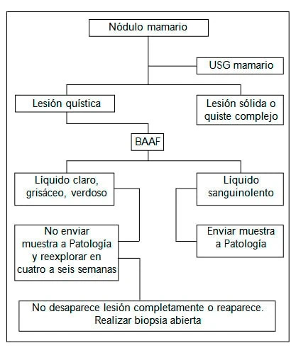
Cystic lesions
If the ultrasound shows a cystic lesion, the following step is a fine needle aspiration biopsy (FNA), because it is a low-cost diagnostic and therapeutic option.
Among false-positive explanations there are interpretation mistakes, inadequate preparation, cell distortion, reactive atypia in mastitis or fat necrosis, post-radiation or post-chemotherapy, as well as epithelial hyperplasia, gestational changes, benign papillary lesions, and sclerosing adenosis
Cancer risk depending on cytology report.
Cytology Malignancy percentage
- Malignant 99.2-100
- Suspicious 50-70
- Benign 0.2-15.2
- Unsatisfactory 5-22
- Undetermined 5-22
Solid lesions
In solid or complex lesions, physical examination, image studies, and the FNA allow for a quick diagnosis without delaying treatment, because together, they have 100% sensitivity and specificity; also, they reduce up to 50% of the costs for each evaluated mass.
Suspicious, probably malignant (the cytologic results are highly suggestive of malignancy).
- Malignant.
- Unsatisfactory (due to scarce cellularity, artifacts due to low cellularity, limited because of blood or inflammatory processes).
The cancer risk level on each category determines the management strategy and the proportion of women with cancer in each of the categories.
Request a Second Medical Opinion
WE WORK FOR YOUR HEALTH
SPECIALIZED TREATMENTS
CONTROL AND DIAGNOSIS
MAXIMUM TECHNOLOGY
Contact Us
Leave us your data and consultation to offer you personalized medical advice
International Consultation
If you want remote medical care, you can consult
through the following links:
International Appointment
Online Consultation
Whatsapp: + 34 679 112 179